Evaluation of cell viability and cytotoxicity using the SpectraMax i3x Multi-Purpose Plate Reader

Take advantage of the SpectraMax i3x multi-function microplate reader
Evaluation of cell viability and cytotoxicity by ultrasensitive chemiluminescence detection
 
Advantage
1. The instrument has ultra-high sensitivity chemiluminescence detection function, as low as 10 cells per hole
2. Microplate reading height is automatically optimized to improve the detection signal strength software preset
3. Software preset template can analyze test results faster
Introduction
The SpectraMax i3x is Molecular Devices' new multi-function microplate reader that uses the chemiluminescence detection capabilities of the instrument for cell viability and cytotoxicity. The instrument can sensitively and quickly detect the number of living cells in the medium and the cytotoxicity after corresponding treatment. Promega's CellTiter-Glo reagent utilizes the ATP participation in the firefly luciferase reaction system to make it emit light. The intensity of the chemiluminescence signal depends on the level of ATP in the medium, that is, it depends on the living cells. How many are the numbers. A biochemical chemiluminescence detection kit based on biochemiluminescence principle from BioVision for the detection of adenylate kinase (AK), a common protein found in all cells, which destroys cell membrane integrity It is released into the medium and AK can convert ADP to ATP, so chemiluminescence detection can be performed in a similar manner.
material
CellTiter-Glo Luminescent Cell Viability Assay (Promega P/N G7570)
. Bioluminescence Cytotoxicity Assay Kit (BioVision P/N K312-500)
HeLa. Cells (ATCC P/N CCL-2)
Black bottom 96-well cell culture plate (Corning P/N 3904)
White 96-well cell culture plate (Corning P/N 3917)
SpectraMax i3x Multi-Purpose Plate Reader
method
Preparation reagent
The CellTiter-Glo buffer and substrate were thawed before use and equilibrated to room temperature. CellTiter-Glo buffer was added to a brown vial containing the CellTiter-Glo substrate and the reagent was gently inverted as indicated in the kit instructions. Mix well. For the biochemiluminescent cytotoxicity test kit, a bottle of AK detection reagent is included, and 1.1 ml of AK reagent buffer is added to the reagent before use and gently mixed, and it is necessary to equilibrate for 15 minutes at room temperature. This 10X AK reagent stock solution needs to be diluted before it becomes a reagent buffer.
Relationship between cell number and chemiluminescence signal
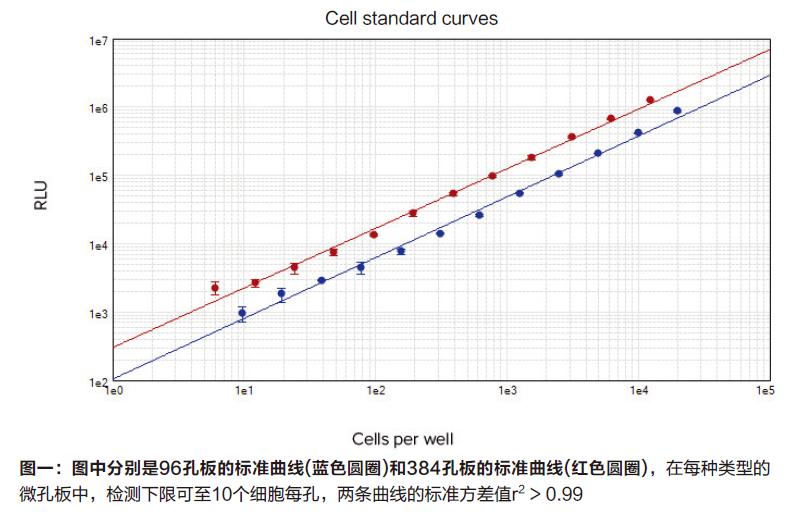
Hela cells were cultured in MEM medium containing 10% fetal calf serum and double antibody, and the cells were trypsinized and suspended in a medium for counting. The treated cells were plated in 96-well plates and diluted from cell culture medium to a density of from 50,000 cells per well / 100 ul to 10 cells per well / 100 ul. If cells are plated in 384-well plates, the density after dilution is from 12,500 cells per well / 25 ul to 6 cells per well / 25 ul. Only the cell culture medium was added to the control wells to detect the chemiluminescent signal values ​​of their background.
During cell testing, the exact number of cells can be used to generate a standard curve, and different volumes of CellTiter-Glo reagent are added to the corresponding well plates, such as 100 ul (96 well plates) or 25 ul (384 well plates). Place the microplate on the shaker for 2 minutes, incubate for 10 minutes at room temperature, and wait until the chemiluminescence signal value is stable before testing.
ATP standard curve
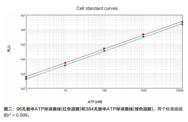
ATP was diluted at a 1:10 dilution and added to 96-well plates and 384-well plates at concentrations ranging from 10 uM to 1 nM, with no cell-filled wells as blank wells. CellTiter-Glo reagents can be used to detect ATP and use the SpectrMax i3x's microplate optimization settings and read height optimization settings to help you get better signal values ​​before testing.
Cell viability and cytotoxicity test
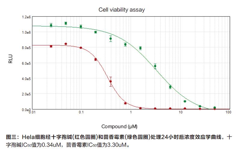
HeLa cells were pre-plated in a white-bottomed cell culture type 96-well plate at a density of 15,000 per well and cultured overnight. On the second day, two compounds, staurosporine and anisomycin, were added to the cell wells to induce apoptosis. Both compounds started at a concentration of 50 uM and were diluted 1:24 to 24 nM. After a 24-hour incubation, the treated cells were assayed using the CellTiter-Glo Cell Viability Assay Kit and BioVision's Cytotoxicity Assay Kit, respectively.
For the CellTiter-Glo cell viability assay, 100 ul of CellTiter-Glo reagent was added to a white microplate containing cells and waited for about 10 minutes. After the cells were completely lysed, the corresponding assay was performed.
For the BioVision cytotoxicity assay, 100 ul of treated cell culture medium was taken from each well, transferred to a white microplate, and 100 ul of AK working solution was added to the white microplate. This method took 30 minutes. Complete the test.
Instrument setup and results analysis
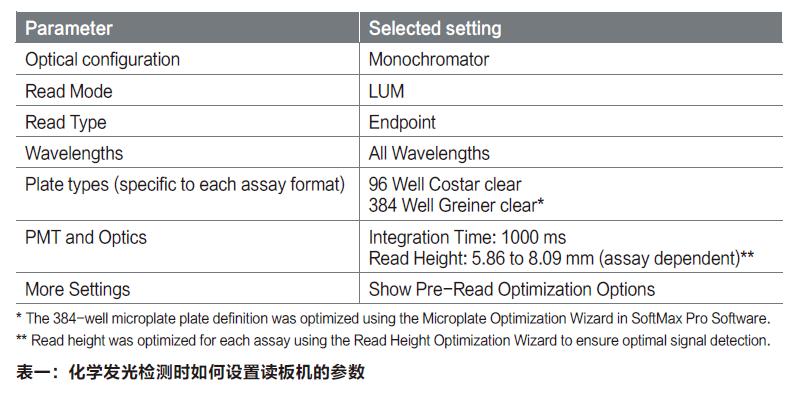
The parameters of the SpectraMax i3x reader are shown in Table 1. The average relative chemiluminescence signal (RLU) and standard deviation values ​​are obtained directly using the professional grade SoftMax. pro software. The cell dilution curve and the ATP standard curve were directly drawn, and the 4-parameter fitting method was selected to fit the IC 50 value. There are also templates for the CellTiter-Glo detection test in the software preset template library that can be easily called directly.
result
When the SpectraMax i3x reader is tested on 96-well and 384-well plates, the minimum detection limit is 10 cells per well. The sensitivity of this assay is much higher than standard colorimetry and most fluorescence methods. The CellTiter-Glo reagent detection linear range covers all cell concentration intervals, with a dynamic range of more than 3 orders of magnitude and r2 > 0.99 (Figure 1).

The ATP standard curve can be used to verify both the test conditions and the intracellular ATP content. Therefore, the ATP standard concentration range from 1 nM to 10 uM produces a range of chemiluminescent signals that cover the signal values ​​generated in the entire number of different cell numbers (Figure 2), with a linear range of detection greater than four orders of magnitude.
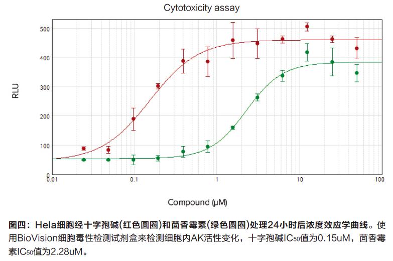
Changes in cell viability after treatment with compounds such as staurosporine and anisomycin can be achieved by detecting ATP levels (Figure 3). Cytotoxicity can be achieved by detecting changes in the amount of AK (Figure 4). Staurosporine (0.34uM and 0.15um) and anisomycin (3.30uM and 2.28uM) twice treated experimentally obtained similar IC 50 values.

in conclusion
The ATP-based cell viability and AK-based cytotoxicity assays were performed with the SpectraMax i3x plate reader chemiluminescence detection function, which showed ultra-high sensitivity with a detection limit of 10 cells per well. A four-parameter fit can be performed with a professional-grade SoftMax Pro software, followed by a direct calculation of the IC50 value. The software automatically optimizes the detection height of the microplate to help the user get the best signal value.
In addition to the chemiluminescence detection function, the SpectraMax i3x reader can be upgraded with more detection functions to meet different detection requirements of different tests. The instrument's light absorption and fluorescence intensity detection features a proprietary spectral fusion technology (Spectra FusionTM Illmumination) that combines wavelength flexibility with optimal detection signals.
The host can be added to the cartridge with the new detection function, and the user can upgrade the function at any time, including time-resolved fluorescence detection and Western Blot detection. The Spectra. MiniMaxTM 300 cell imaging module can also be selected to extend the reader's detection capability. And the scope of application. All functions of the instrument can be operated with one software, namely the professional level SoftMax Pro software, which includes nearly 120 preset detection templates such as CellTiter-Glo, which is convenient for users to quickly collect data and analyze data.

Active Pharmaceutical Ingredients

Active Pharmaceutical Ingredients(API) refer to the raw materials used in the production of various preparations. They are the effective ingredients in the preparations. They are various powders, crystals, extracts, etc., prepared by chemical synthesis, plant extraction or biotechnology, but Substances that the patient cannot take directly. API is intended to be used in any substance or mixture of substances in the manufacture of pharmaceuticals, and when used in pharmaceuticals, it becomes an active ingredient of the pharmaceuticals. Such substances have pharmacological activity or other direct effects in the diagnosis, treatment, symptom relief, treatment or prevention of diseases, or can affect the function or structure of the body. According to its source, active pharmaceutical ingredients are divided into two categories: synthetic chemical active Pharmaceutical ingredients and natural chemical active Pharmaceutical ingredients.

Chromium Picolinate,Tianeptine,6-Paradol,Aminobutyric acid,acetylcysteine,L-Carnosine

Xi'an Gawen Biotechnology Co., Ltd , https://www.ahualynbio.com